Systematic Review of Mindfulness Induced Neuroplasticity in Adults: Potential Areas of Interest for the Maturing Adolescent Brain
Virginie Kirk, Charikleia Fatola and Marina Romero Gonzalez
DOI10.4172/2472-1786.100016
Institute of Psychiatry Psychology and Neuroscience, King’s College, London,UK
- *Corresponding Author:
- Virginie Kirk
Child and Adolescent Mental Health Student, Institute of Psychiatry Psychology and Neuroscience, King’s College, London, SE5 8AF, United Kingdom.
Tel: +447838169923
E-mail: virginie.kirk@kcl.ac.uk
Received Date: November 04, 2015; Accepted Date: February 08, 2016; Published Date: February 22, 2016
Citation: Kirk V, Fatola C, Gonzalez MR. Systematic Review of Mindfulness Induced Neuroplasticity in Adults: Potential Areas of Interest for the Maturing Adolescent Brain. J Child Dev Disord. 2016, 2:1.doi: 10.4172/2472-1786.100016
Abstract
We present a systematic review of studies documenting neural functional alterations after mindfulness training in adult samples. Adolescence is a critical developmental period characterised by the late maturation of the prefrontal cortex and functional stabilisation, processes that are both genetically led and experience dependent. Thus adolescence could be an opportunity to redress the developmental trajectory. Adjustment problems may evolve into maladaptive behaviours and psychological disorders. This is an important public health concern that justifies investing in adolescent focused research. Mindfulness has received increasing attention over the last thirty years, for its clinical and protective effects. The present review focuses on the wider effects of mindful attention on functional processes. Our results indicate that mindful attention practices are associated with extensive functional plasticity. Reduced activation has been observed in the limbic system and the default mode network. In contrast, increased engagement of executive and attention related areas have been observed in the anterior cingulate cortex, the ventro- and dorso-medial prefrontal cortex. As the limbic system, default mode network and executive functions all undergo major maturation processes in adolescence, research is needed to understand how mindful attention could contribute, to the experience driven plasticity of the emergent functional systems associated to self-referential, socio-emotional stress and higher executive capabilities in adolescents.
Introduction
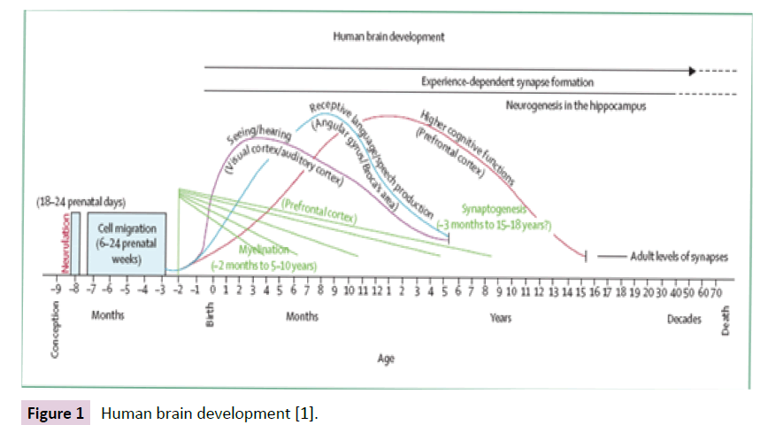
Adolescence is a critical period of development, taking place in the context of major social, physical, and psychological change. The late maturation of prefrontal regions is associated with a shift from the plasticity of early life towards stability. Transformations include an initial proliferation followed by gradual decline in the production and pruning of synaptic connections, processes that are both genetically driven and experience dependent (Figure 1) [1]. These affect higher order executive functions, inhibitory control, planning, emotional regulation and functional connectivity within distal regions [2,3]. Puberty hormonal changes have been associated to dramatic functional and structural changes [4], with increased reactivity to social and emotional information [5]. The relative dominance of affective over executive systems is thought to be responsible for higher risk taking behaviour. In this context, adjustment problems may evolve into maladaptive behaviour and the onset of psychological disorders [2]. Preventive care would be welcome.
Based on Buddhist meditative practices, mindfulness brings awareness to the current moment experience. Mindfulness has been conceptualised as having three components: first, practices developing the self-regulation of attention; second, developing an attitude of openness, curiosity and acceptance toward one’s experience; third, developing intentional non-judgemental focus in the present as it unfolds [6]. Meditative practice aims to develop mindful attention. This in turn develops greater mindfulness awareness. During practice, one can chose to deploy mindful attention to different objects of their current inner and outer experience, such as breathing, sensations felt in the body (bodyscape), noises heard in the environment (soundscape), thoughts and feelings coming and going into the mind (mindscape) or even walking. For a full review of practices, please refer to Kabat-Zinn. This review is concerned with mindful attention to the breath or physical perception. This involves observing the flow of air entering and leaving the body, the physical sensations associated with breathing as the deliberate focus of attention and an anchor into the present moment. When attention drifts away from breathing, noticing that attention has wandered away into thoughts and bringing attention back to the breath; accepting that attention gets distracted; acknowledging our reaction when this happens and letting go of it without judging; and kindly returning attention back to breathing [7].
Mindfulness has been attracting much support in recent years. It was integrated into Western clinical practice in the form of Mindfulness Based Stress Reduction (MBSR) [8], which has inspired other mindfulness-based interventions including depression relapse prevention (Mindfulness Based Cognitive Therapy, MBCT) [9], pain management [10], smoking cessation [11] and addiction relapse [12]. Another interesting aspect of mindfulness is its preventive health-promoting stress-reducing capacity in non-clinical [13, 14] and in adolescent populations using school based delivery models [15]. For a full review of school programmes, please refer to Zenner and colleagues.
MBSR and other mindfulness-based programs have proved effective at treating stress related and other clinical disorders [7]. However the neural mechanisms of mindfulness are still poorly understood. This has inspired a new interdisciplinary field of research, mindfulness neuroscience, seeking to explore the neural mechanisms associated with mindfulness practice [16]. Two key beneficial areas have been highlighted: one, the effect of mindfulness on attention processes; second, mindfulness as an emotion regulation strategy, exemplified by the emotional stability that characterises long-term meditators. Chiesa and colleagues’ review proposed that mindfulness involves two kinds of emotion regulation processes: top-down regulation of prefrontal regions over the limbic system in novice practitioners, and reduced reactivity bottom-up process in experienced meditators.
There are a number of areas of the adult brain that have been identified and associated with the practice of mindfulness
The limbic system, sometimes referred to as the emotional brain, comprises a number of structures working together and involved in emotions, motivation and long-term memory. In particular the amygdala has a primary role in the recognition of fear, facial emotions and emotional reactivity. Connected to the autonomous system, it prepares the body for a fight or flight response [17].
The default mode network (DMN) is a network of areas within the medial prefrontal cortex, including the medial temporal lobes, posterior cingulate and medial temporal cortices. It is associated with episodic memory, introspection and self-referential processing. It is most active when the brain is not engaged in any specific task. Connectivity within the DMN is thought to reflect mind wandering [18]. Activity in the DMN has been associated with thought rumination, a feature strongly associated with depressive disorders [9]. There is limited evidence of the DMN in the infant brain. Connectivity within the DMN becomes more consistent in children aged nine to twelve years, suggesting developmental change [19].
The right ventrolateral prefrontal cortex (vlPFC) is a component of the attention network governing reflexive reorienting. The vlPFC is thought to modulate emotional affect on executive functions and fully matures in late adolescence [19].
The dorsolateral PFC (dlPFC) is associated with executive functions such as working memory, vigilance and sustained attention but also planning, inhibition, abstract reasoning and risky decisionmaking. It is well interconnected to attention related areas [19].
The insula (INS) is thought to map the body states associated with emotions giving rise to conscious feelings, providing emotional context to sensory experience. Receiving homeostatic input from the thalamus and sending output to the limbic system it is involved in homeostasis, perception, motor control, self-awareness, cognitive functioning and interpersonal experience [20].
The anterior cingulate cortex (ACC) is thought to act as an interface between the limbic and frontal regions, between emotions and cognition, converting feelings into intention and action, controlling emotions, concentrating on problem solving and making adaptive responses to the environment. The dorsal part (dACC) is a central station for processing top-down and bottom-up stimuli, and assigning control to other areas. The ventral part of the ACC (vACC) is connected with the limbic system and anterior insula, and involved in assessing the salience of emotion and motivational information [19, 20].
This review proposes to revisit neuroimaging studies of mindfulness over the last ten years, and the central role to mindful attention practice, to tease out its impact on the functional activity of the brain, and how it could relate to maturation processes of the adolescent brain.
Methods
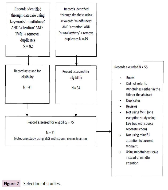
The selection process of the articles included in this review can be visualised in Figure 2. Functional imaging studies were gathered from a comprehensive database search in November 2014, using the Ovid platform, including Embase, PubMed, Medline, PsychInfo, PsychArticles, and Ovid Full Text Journals. A first string of keywords combined ‘mindfulness’ AND ‘attention’ AND ‘fMRI’ and ‘remove duplicates’. Another string combined ‘mindfulness’ AND ‘attention’ and ‘neural activity’ and ‘remove duplicates’. A first screening removed all studies that did not refer specifically to mindfulness either in the title or the abstract. A second screening removed irrelevant studies and duplicates between the two searches.
Inclusion criteria included: neuroimaging studies of the effects of mindfulness captured as mindful attention, using fMRI measure. Exclusion criteria were as follow: studies that did not refer specifically to mindfulness either in the title or in the abstract, studies that did not use neuro-imaging methods, studies based on mindfulness rating scales instead of mindful attention practice. Most studies were based on mixed adult population samples, some with a brief induction to mindful attention, some following an eight-week training programme, some having several years of daily practice. Of note, there were no published fMRI studies available using children or adolescent samples.
Results
Previous reviews [7, 21] have emphasized the challenge of capturing ‘mindfulness’ as a variable. To resolve this difficulty, this review selected articles that captured mindfulness as mindful attention. As noted earlier, during mindfulness practice, one can chose to deploy mindful attention to varying aspects of their current moment experience. Objects of mindful attention may include breathing, body sensations, sounds in the environment, thoughts and feelings. In the studies selected for this review, independently of their level of training and experience, participants were asked to practice mindful attention while completing an experimental task in the scanner. Controls usually performed the same task without mindful attention. Details of the results of individual studies selected are available in Appendix 1.
Deactivation of the limbic system
Studies have associated mindful attention with deactivation of the limbic system, particularly in the amygdala [22-25], after only one week training [26], and a negative correlation between practice and activation [26, 27]. Weaker signals in the thalamus suggested higher arousal and vigilance, in experts compared to novices [28].
Additionally articles reported lower resting states in the amygdala in experts [14] but also higher activation in experts [22]. This discrepancy could be explained by differences in response, with an initial spike, followed by faster return to baseline resting state, suggesting higher initial neurobiological reactivity but faster extinction of emotional responses in experts [29].
Deactivation of the cortical midline
Articles reported deactivation of sub-regions along the cortical midline [30], reduced signals in the ventromedial prefrontal cortex (vmPFC) [25] and dorsomedial prefrontal cortex (dmPFC) [29]. Bilateral dmPFC deactivation was concomitant with bilateral activation in rostral ACC [31]. Deactivation [24] of those subregions of the mPFC has been associated with a shift of activation in right lateralised PFC and para-limbic structures [24].
Deactivation of the default mode network
Articles reported deactivation in the DMN [25, 32], reduced signal in the DMN at baseline in novices in mindful focus group [24], deactivation of DMN regions after only one week training, along with reduced interconnectivity within DMN regions in experts compared to novices, suggesting that expertise consolidates this deactivation [26].
Reduced recruitment and interconnectivity within the DMN sub-regions has been associated with greater connectivity from DMN to sensory cortices [18], executive and salience network, ACC, INS and thalamus [25], and inversely correlated to the dlPFC recruitment [22].
One study, however, reported stronger connectivity between DMN regions and right IPL, suggesting significant differences between experts and novices [26].
Executive and attention related areas in the prefrontal cortex
Mindful attention has been associated with increased engagement of prefrontal areas [23]. Indeed articles have reported: activation in visual attention [29]; greater recruitment of the right INS, right subgenual ACC, vlPFC, related to volitional orienting [33]; increased activation of bilateral central executive regions, dlPFC, during the anticipation of pain [22, 34]; increased activation in dlPFC, vlPFC and right lateral IPC, when shifting attention back to breathing and increased activation of the dlPFC while maintaining focused attention [35].
Articles reported widespread prefrontal activation during affect labelling, associated with simultaneous bilateral amygdala deactivation [23]. Post-training, activation in visual attention was associated with deactivation in right amygdala [29] suggesting a top down inhibition of limbic responses [23]. Prefrontal activation in the dlPFC and vlPFC was also associated with deactivation in the DMN, during experiential focus and without previous training [24]. The engagement of prefrontal areas seems concomitant to the disengagement of limbic and DMN systems.
However, a study of experienced meditators, attending to pain, reported a decrease in signal in the right vlPFC, dlPFC, mPFC, and orbitofrontal cortex along with increased signal in posterior parietal attention related regions [25], suggesting lesser engagement of prefrontal regions with experience in response to pain.
The insula and the anterior cingulate cortex: interoceptive awareness and executive salience
Articles have reported increased signal in the insula, in relation to interoceptive awareness, increased signal post-training, during a subjective appraisal task [24], increased signal propagation between the posterior and anterior INS, and reduced connectivity between the INS and dmPFC [36], suggesting changes in balance between interoceptive attention and external exploration [36].
Articles have reported the simultaneous recruitment of the ACC and the INS [32] greater ACC activation in novices, consistent with its role in learning and conflict monitoring [27], and signal increase in the bilateral anterior INS and dACC during awareness of mind wandering, consistent with conflict monitoring [35], Stronger connectivity between the left INS and dACC, in response to pain in experts suggested increased viscera-somatic awareness and lower pain related responses in primary affective sensory areas [25]. Increased engagement of the dACC, INS and thalamus, was associated with the disengagement of the DMN, suggesting a shift of executive salience network over self-referential processing. Monitoring experience via the dACC may illuminate and extinguish conditioned responses [25].
One study however reported deactivation in interoception related areas in the cortical midline with simultaneous signal increase in right PCC, suggesting a spike in activity followed by rapid decrease and underscoring the importance of the timing in measures [37] (Table 1).
Deactivation in the limbic system
| Amygdala | Deactivation | Brown et al. [22] Creswell et al.[23] Farbet al.[24] Grant et al.[25] |
| After only one week training | Taylor et al.[26] | |
| Negative correlation between greater practice and activation | Brefczynski-Lewis et al.[27] Taylor et al. [26] |
|
| Lower resting state in experts | Jhaet al.[14] | |
| Higher activation in experts | Brown et al.[22] | |
| Faster extinction in experts | Goldinet al. [29] | |
| Thalamus | Weaker signal in experts vs novices, suggesting higher arousal | Lee et al.[28] |
Deactivation in the cortical midline
| Sub-regions | Reduced activation of sub-regions | Grant et al.[25] Zeidanet al.[30] |
| Associated to shift of activation in right lateralised PFC | Farbet al.[24] | |
| vmPFC | Reduced signal | Grant et al.[25] |
| dmPFC | Deactivation in dmPFC | Goldinet al. [29] |
| Bilateral deactivation concomitant with bilateral ACC activation | Holzelet al. [31] |
Deactivation in the default mode network
| DMN | Deactivation of sub-regions | Dickenson et al.[32] Grant et al.[25] |
| Reduced signal at baseline in novices in mindful attention | Farbet al.[24] | |
| Deactivation after only one week training | Taylor et al.[26] | |
| Reduced interconnectivity within sub-regions in experts | Taylor et al.[26] | |
| Expertise consolidates deactivation | Taylor et al.[26] | |
| Reduced recruitment and interconnectivity associated with greater connectivity from DMN to sensory cortices | Kilpatrick et al.[18] | |
| Reduced recruitment and interconnectivity associated with greater connectivity from DMN to executive and salience networks (ACC, INS and thalamus) | Grant et al.[25] | |
| Inversely correlated to dlPFC recruitment | Brown et al. [22] | |
| Stronger connectivity between DMN regions and right IPL, suggesting differences between experts and novices | Taylor et al.[26] |
Recruitment of executive and attention related areas in the prefrontal cortex
| PFC | Diffuse engagement associated to bilateral amygdala deactivation | Creswellet al.[23] |
| vlPFC, | Recruitment related to volitional orienting | Farbet al.[33] |
| dlPFC | Bilateral activation | Brown et al.[22] Way et al.[34] |
| dlPFC | Increased activation while sustaining attention | Hasenkampet al.[35] |
| dlPFC, vlPFC | Simultaneous activation with inferior parietal cortices when shifting attention back to breathing | Hasenkampet al. [35] |
| Associated to deactivation of the DMN | Farbet al.[24] | |
| Decreased engagement in right vlPFC, dlPFC and mPFC in experienced meditators in response to pain | Grant et al.[25] |
Insula and anterior cingulate cortex: interoceptive awareness and salience
| Insula | Increased signal post training | Farbet al.[24] |
| Increased signal propagation between posterior & anterior INS | Farbet al.[33] | |
| Reduced connectivity with dmPFC | Farbet al.[33] | |
| Simultaneous recruitment of the ACC and the INS | Dickenson et al.[32] | |
| ACC | Greater activation in novices (conflict monitoring) | Brefczynski-Lewis et al.[27] |
| Increase in bilateral anterior INS and dACC | Hasenkamp et al. [35] | |
| Stronger connectivity between left INS &dACC in experts | Grant et al.[25] | |
| Engagement of dACC, INS & thalamus associated with disengagement of the DMN | Grant et al.[25] |
Table 1: Summary of results.
Discussion
Summary of results
Mindful attention whilst carrying out an experimental task in the scanner is associated with a disengagement of the limbic system, particularly the amygdala, a reduction of its resting state in expert meditators and reduced emotional reactivity. Results suggested a disengagement of regions in the cortical midline, particularly the DMN, and reduction of connectivity within DMN sub-regions suggesting less mind wandering, and reduced connectivity with other networks suggesting less engagement in self-referential processes. Simultaneously to the deactivation of the limbic system and the DMN, results suggested an increased engagement of the INS and ACC, areas related to viscera-somatic awareness and salience, suggesting a move away from affective to interoceptive monitoring [33]. Importantly, results suggested an increased engagement in the prefrontal cortex of areas involved in executive attentional processes. These underscore important functional plasticity, a shift away from emotional reactivity and self-referential processes towards interoceptive and perceptual awareness with greater attention allocation.
Strengths of this review
This review highlights that mindful attention, independently of experience, induces functional neuroplasticity and identifies the functional systems that are modified by this practice. In contrast to previous work, we were concerned specifically with the effects of mindful attention on brain circuitry. Our analysis indicates that staying on task requires the recruitment of attentional resources, triggering a cascade of functional alterations. Resources are allocated to focus, maintain, monitor, resolve conflict and re-orient attention back to the task, every time the mind wanders away. Working memory is also involved in maintaining attention [14].
This review underscores how the global recruitment of attentional resources, PFC, dlPFC, vlPFC and OFC [23], deactivates the limbic system [26], in turn disengaging automatic responses and habitual reactivity, with increased resources allocated to attentional processes to attended and unattended information within and across sensory modalities [18]. Thus emotional regulation may be a by-product of mindful attention, as resources are redirected away from the limbic areas and the DMN. This deactivation may be intrinsic to mindful attention and not limited to experienced individuals. Controlling mind wandering and suspending habitual automatic responses promotes present moment conscious awareness [7].
Implications for the adolescent brain
Mindful attention appears to redistribute resources between the DMN, limbic system and executive functions. These undergo important maturational changes in adolescence, with region specific alterations in the amygdala, and delayed maturation of top-down control systems in the prefrontal cortex, attention regulation and response inhibition [2].
First, there is limited evidence of the DMN in the infant brain. Connectivity within the DMN becomes more consistent in children aged nine to twelve years, suggesting an important developmental window [19]. As the DMN is involved in selfreferential episodic memory and has been linked to a ruminative thinking style, itself linked to depressive disorders, it would be interesting to find out how mindful practice can support the consolidation of this network in adolescence and prevent the onset of depressive disorders in those at risk.Second, research suggests higher sensitivity to stress and limbic reactivity, exaggerated startle response in fear processing and stronger interference of emotional stimuli on task completion [38] and heightened autonomic responses, in cortisol measure, during challenging situations [39]. An over-activated stress system may potentially damage lifelong learning and memory consolidation [40]. Thus adolescence is a particularly sensitive period for stress and the onset of anxiety disorders [15]. On the basis that mindful attention practice in adults alters limbic responses, it would be worth investigating how mindful practice could benefit the maturation of the limbic system and regulate stress reactivity in adolescence.
Third, adolescents process information differently to prepubertal children and adults. With enhanced reactivity to social stimuli [5], immature cognitive controls can be overwhelmed. The challenging conditions in adolescence of increased task demand and heightened emotional arousal can reduce decision making ability, a phenomenon known as hot cognition, and thought to be responsible for engaging in risky behaviour [41]. This could be an area of potential benefice for adolescents, as mindful attention has been shown in adults to engage executive prefrontal areas, redirecting resources away from automatic conditioned responses [7].
The adolescent brain plasticity is an important window of opportunity when critical experiences can still alter the developmental trajectory [2]. We argue that mindful attention practices could enhance adolescent brain plasticity, and promote adolescents’ resilience to stress, anxiety, depression and poor decision-making whilst boosting the maturation of prefrontal regions.
Limitations
There were notable divergences between studies particularly regarding the engagement of the ACC. Discrepancies can be explained in part by methodological limitations. Studies have employed tasks as diverse as attending to pain which involves managing the anticipation of fear, listening to sounds in the scanner which involves attending to the exterior environment or affect labelling which involves subjective referential processes. Each study eliciting different neural responses, and comparing them to different control tasks may affect results. To that effect, studies were selected using mindful attention as a constant variable. Second, fMRI measure of BOLD signals include artefact and cannot differentiate between inhibitory or excitatory neural activation [12]. This make the interpretation of signals difficult, as increased activity in one area could reflect either inhibition or excitation processes. However in most studies additional behavioural data has been used to interpret results and the direction of change [12]. Importantly, many studies have employed a cross-sectional design, which prevents the establishment of a causal relationship between the observed effects and mindfulness practice. Nevertheless, longitudinal studies, have reported increased cortical thickness with pervasive grey matter changes, mirroring changes reported in cross-sectional studies and supporting the idea that long term practice is associated with neuroplasticity [31].
Finally the case of the ACC differential engagement, suggests different processing between novices and experienced practitioners. This raises the questions of when process changes take place, when practice solidifies [14], how much practice is required for changes to occur [7], whether effects can transfer to non-mindful states, and do effects translate into enduring changes in mental functions.
Conclusion and recommendations for future research
Further research into adolescent brain maturational processes is needed not from a public health but also humanistic perspective. Future research could identify periods of optimal plasticity for different functional systems and how mindful attention could enhance this plasticity, to optimise or redress the adolescent developmental trajectory.
In their review of preliminary studies, Zenner and colleagues concluded that school can be an appropriate setting for carrying out research, as mindfulness interventions are well suited to group delivery. They highlighted that school settings can facilitate the delivery of eight-week mindfulness programs adapted to suit the requirements of adolescents. Such programs were well received by both teachers and students. Their review concluded that mindfulness practice could be integrated into the curriculum as a low cost universal prevention program [42]. Thus we recommend that a study could be organised to explore the effect of an eight-week school based mindfulness program, at different ages across adolescence, with fMRI measures taken at baseline and post program.
References
- Thompson RA, Nelson CA (2001) Developmental science and the media: Early brain development. ?Am. Psychol 56: 5.
- Spear LP (2013) Adolescent neurodevelopment. J Adolesc Health 52: S7-S13.
- Giedd JN, Blumenthal J, Jeffries NO, Castellanos FX, Liu H, et al. (1999) Brain development during childhood and adolescence: a longitudinal MRI study. Nat Neurosci 2: 861-863.
- Romeo RD (2010) Adolescence: a central event in shaping stress reactivity. DevPsychobiol52: 244-253.
- Blakemore SJ (2008) Development of the social brain during adolescence. Q J ExpPsychol 61: 40-49.
- Shapiro S L, Carlson LE, Astin, JA, Freedman B (2006) Mechanisms of mindfulness. J ClinPsychol62: 373-386.
- Williams JMG (2010) Mindfulness and psychological process. Emotion 10: 1.
- Kabat-Zinn J (1994) Wherever you go, there you are: Mindfulness meditation in everyday life: Hyperion.
- Segal ZV, Williams JMG, Teasdale JD (2012) Mindfulness-based cognitive therapy for depression: Guilford Press.
- Burch V, Penman D (2013) Mindfulness for Health: A practical guide to relieving pain, reducing stress and restoring wellbeing: Little Brown Book Group.
- Westbrook C, Creswell JD, Tabibnia G, Julson E, Kober H, et al. (2013) Mindful attention reduces neural and self-reported cue-induced craving in smokers. SocCogn Affect Neurosci8: 73-84.
- Witkiewitz K, Lustyk MKB, Bowen S (2013) Retraining the addicted brain: A review of hypothesized neurobiological mechanisms of mindfulness-based relapse prevention. Psychol Addict Behav. 27: 351.
- Ma SH, Teasdale JD (2004) Mindfulness-based cognitive therapy for depression: replication and exploration of differential relapse prevention effects. J Consult ClinPsychol 72: 31-40.
- Jha AP, Stanley EA,Kiyonaga A, Wong L, Gelfand L, et al. (2010) Examining the protective effects of mindfulness training on working memory capacity and affective experience. Emotion 10: 54.
- Broderick PC, Jennings PA (2012) Mindfulness for adolescents: A promising approach to supporting emotion regulation and preventing risky behavior. New Dir Youth Dev136: 111-126.
- Tang YY, Posner MI (2012) Special issue on mindfulness neuroscience. SocCogn Affect Neurosci nss104.
- LeDoux JE, Iwata J, Cicchetti P, Reis DJ (1988) Different projections of the central amygdaloid nucleus mediate autonomic and behavioral correlates of conditioned fear. J Neurosci 8: 2517-2529.
- Kilpatrick LA, Suyenobu BY, Smith SR, Bueller JA, Goodman T, et al. (2011) Impact of mindfulness-based stress reduction training on intrinsic brain connectivity. Neuroimage56: 290-298.
- Broyd SJ, Demanuele C, Debener S, Helps SK, James CJ, et al. (2009). Default-mode brain dysfunction in mental disorders: a systematic review. NeurosciBiobehav Rev 33: 279-296.
- Gu, X, Hof PR, Friston KJ, Fan J (2013) Anterior insular cortex and emotional awareness. J Comp Neurol 521: 3371-3388.
- Chiesa A, Serretti A, Jakobsen JC (2013) Mindfulness: Top–down or bottom–up emotion regulation strategy? ClinPsychol Rev 33: 82-96.
- Brown CA, Jones AKP (2013) Psychobiological Correlates of Improved Mental Health in Patients With Musculoskeletal Pain After a Mindfulness-based Pain Management Program. Clin J Pain 29: 233-244.
- Creswell JD, Way BM, Eisenberger NI, Lieberman MD (2007)Neural correlates of dispositional mindfulness during affect labeling. Psychosom Med 69: 560-565.
- Farb NA, Segal ZV, Mayberg H, Bean J, McKeon, et al. (2007) Attending to the present: mindfulness meditation reveals distinct neural modes of self-reference. SocCogn Affect Neurosci 2: 313-322.
- Grant JA, Courtemanche J, Rainville P (2011)A non-elaborative mental stance and decoupling of executive and pain-related cortices predicts low pain sensitivity in Zen meditators. PAIN 152: 150-156.
- Taylor VA, Daneault V, Grant J, Scavone G, Breton E, et al. (2013) Impact of meditation training on the default mode network during a restful state. SocCogn Affect Neurosci8: 4-14.
- Brefczynski-Lewis JA, Lutz A, Schaefer HS, Levinson DB, Davidson R J, et al. (2007) Neural correlates of attentional expertise in long-term meditation practitioners. ProcNatlAcadSci 104: 11483-11488.
- Lee TM, Leung MK, Hou WK, Tang JC, Yin J, etal. (2012) Distinct neural activity associated with focused-attention meditation and loving-kindness meditation. Plus One.
- Goldin PR., Gross JJ (2010) Effects of mindfulness-based stress reduction (MBSR) on emotion regulation in social anxiety disorder. Emotion 10: 83.
- Zeidan F, Martucci KT, Kraft RA, Gordon NS, McHaffie JG, et al. (2011) Brain mechanisms supporting the modulation of pain by mindfulness meditation. ?J. Neurosci31: 5540-5548.
- Hölzel BK, Ott U, Hempel H, Hackl, A, Wolf K, et al. (2007) Differential engagement of anterior cingulate and adjacent medial frontal cortex in adept meditators and non-meditators. ?NeurosciLett421: 16-21.
- Dickenson J, Berkman ET, Arch J, Lieberman MD (2012)Neural correlates of focused attention during a brief mindfulness induction. SocCogn Affect Neuroscinss030.
- Farb NA, Anderson A K, Mayberg H, Bean J, McKeon D, et al. (2010). Minding one’s emotions: mindfulness training alters the neural expression of sadness. Emotion 10: 25.
- Way BM (2010) Dispositional mindfulness and depressive symptomatology: correlations with limbic and self-referential neural activity during rest. Emotion 10: 12.
- Hasenkamp W, Barsalou LW (2012) Effects of meditation experience on functional connectivity of distributed brain networks. Front Hum Neurosci 1: 6-38.
- Farb NA, Segal ZV, Anderson AK (2012) Mindfulness meditation training alters cortical representations of interoceptive attention. SocCogn Affect Neurosci 8: 15-26.
- Ives-Deliperi VL,Solms M, Meintjes EM (2011) The neural substrates of mindfulness: an fMRI investigation. SocNeurosci6: 231-242.
- Silk JS, Siegle GJ, Whalen DJ, OstapenkoLJ, Ladouceur CD, et al. (2009) Pubertal changes in emotional information processing: pupillary, behavioral, and subjective evidence during emotional word identification. DevPsychopathol 21: 7-26.
- Sumter SR, Bokhorst CL, Miers AC, Van Pelt J, Westenberg PM, et al. (2010) Age and puberty differences in stress responses during a public speaking task: do adolescents grow more sensitive to social evaluation? Psychoneuroendocrinology 35: 1510-1516.
- Andersen SL,TeicherMH (2008) Stress, sensitive periods and maturational events in adolescent depression. Trends Neurosci 31: 183-191.
- Steinberg L (2008) A Social Neuroscience Perspective on Adolescent Risk-Taking. Dev Rev28: 78-106.
- Zenner C, Herrnleben-Kurz, S, Walach H (2014) Mindfulness-based interventions in schools-a systematic review and meta-analysis. FrontPsychol5.
Open Access Journals
- Aquaculture & Veterinary Science
- Chemistry & Chemical Sciences
- Clinical Sciences
- Engineering
- General Science
- Genetics & Molecular Biology
- Health Care & Nursing
- Immunology & Microbiology
- Materials Science
- Mathematics & Physics
- Medical Sciences
- Neurology & Psychiatry
- Oncology & Cancer Science
- Pharmaceutical Sciences


